Thursday, August 26, 2010

How to Use a Journal Article to Advertise Your Product

From this Month's JAMA (1) "Cognitive Behavioral Therapy vs Relaxation With Educational Support for Medication-Treated Adults With ADHD and Persistent Symptoms" by Safren et al.

In order to use an article to advertise your product, first you'll need to chose a topic about which people are fairly ignorant and make yourself an expert on it:

Approximately 4.4% of adults in the United States have attention-deficit/hyperactivity disorder (ADHD).”

Writing Style Recommendation: If you use the word “approximately,” which means “near or around,” then don’t use a number with a decimal. That’s too much precision. Just say “approximately 4% of adults.”

The lead author is Steven Safren, Ph.D., ABPP. In case you’re wondering, those initials mean people have to refer to him as “doctor” and believe that he actually knows what he is talking about. Although adult ADHD is rather amorphous, Safren et al. have extensively researched CBT with adult ADHD. This gives Safren et al. a posteriori authority on the topic.

Second, you'll need to concoct a reason why you're conducting research:

…Medications have been the primary treatment; however, many adults with ADHD cannot or will not take medications while others show a poor medication response. Furthermore, those considered responders to medications may continue to experience significant and impairing symptoms. Thus, there is a need for alternative and next step strategies.

Reader Advisory: Don’t be fooled by this “need.” It’s not a real need, it’s a constructed need (hey look at this huge hole in the ground I created! It needs to be filled!). These authors are using the construct of ADHD and the failure of medication (to treat an arbitrary construct) to accomplish their own goal.

You can read the results in the abstract here (1) as they are only of secondary importance.

Here is the real importance of this article: “Cognitive behavioral therapy was delivered consistent with our manuals.”

The authors of this article are also the authors of these two books (2, 3). This journal article will serve as a marketing device for these manuals.

It works like this: First, they will give “talks” touting the results of this research.

I am going to show you a bunch of statistics that have no real world relevance…

Second, they will conclude that pointless discussion with…

Our manual is available in the literature section located in the lobby.”

Unfortunately, those manuals don’t contain actual cognitive and behavioral material. Sure, they use the jargon, but it’s not actual CBT.

Writing Style Recommendation: Add words to mundane terms, thus making them appear more important than they actually are.

Sessions were designed specifically to meet the needs of ADHD patients and included things like starting and maintaining calendar and task list systems

Anybody can use a calendar or task list, but only “doctors” can provide instruction on how to use a calendar system or a task list system.

Here's a beauty...

The first module focused on psycho-education about ADHD…

What’s the difference between psycho-education and regular education you ask?

If you read an ADHD article on webMD, that’s “education.” When a psychologist prints out that webMD article and hands it to you, that’s “psycho-education.”

Study CBT protocol (i.e., the important one)


Study Relaxation protocol (i.e., the unimportant one)


Other cutting edge cognitive behavioral techniques include “setting priorities” and “breaking large tasks into manageable steps.” Oh snap!

I argue that this is not CBT but rather a collection of common sense solutions to everyday organizational needs. However, Safren et al. refer to this as a particular “type of cognitive behavioral therapy,” which was successfully documented to be useful “as a next-step strategy for patients with ADHD…”

So you better go buy those books!

ResearchBlogging.org

Safren SA, Sprich S, Mimiaga MJ, Surman C, Knouse L, Groves M, & Otto MW (2010). Cognitive Behavioral Therapy vs Relaxation With Educational Support for Medication-Treated Adults With ADHD and Persistent Symptoms: A Randomized Controlled Trial. JAMA : the journal of the American Medical Association, 304 (8), 875-80 PMID: 20736471

Friday, May 28, 2010

Cerebellar Agenesis: Life without a Cerebellum

Many people are familiar with the famous patient H.M., the man who, in an attempt to control his intractable epilepsy, underwent surgical resection of both his medial temporal lobes.

There is another patient who is less famous, known the by initials H.C. He died in 1939 when H.M. was just entering adolescence. Unlike H.M., this patient did not undergo radical resection surgery. In fact, he never underwent brain surgery at all. His contribution to neurology did not begin until after his death at the age of 76.

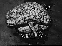
In what was supposed to have been a routine autopsy, H.C. was discovered to have had no cerebellum (see pictured brain above). H.C. had a very rare neurological condition known as cerebellar agenesis. When I was in graduate school, I was taught that the neuroplasticity of the brain was so remarkable, that even a child born without his or her cerebellum could grow-up to have no deficits and live a normal life. As it turns out, that was only partly true. H.C. was in fact, not without deficits. His tale is recounted briefly in two articles from the March 2010 issue of Brain:
"It was clear that there were indeed clinical signs included right external strabismus (i.e., misaligned eyes), slow and slurred articulation and an unsteady gait." (1)
However, he did live a "normal life":
H.C. "had employment, that he was able to work in a manual job and that his working life was not curtailed by his cerebellar agenesis." (1)
What is interesting about H.C. compared to H.M., is that H.C.'s agenesis was discovered only after he died. Very little clinical history about his life exists, making this story a great neurological detective case. Most of what is known about H.C. comes from hospital notes during his last years of life, just before he developed dementia:
"The social history describes him as 'single.' The notes contained a record of his assessment by a neurologist, Dr. Jacobson, who described him as 'a simple man with some hearing loss and slow slurred speech; he has a fair memory for recent and remote events concerning himself, but with limited general knowledge. There is no hallucination or delusions nor emotional defect. He is clean in his habits and able to attend to his person. He is able to get around unassisted." (1)
The human brain is estimated to have approximately 85 billion neurons (2). The cerebellum, which is typically 1/4 the size of the rest of brain, contains a full 50% of all our neurons. If you pay close attention to the image above, you will notice that, in addition to not having a cerebellum, H.C. was also missing his pons, the bulbous structure that is typically adjacent to the cerebellum and is responsible for arousal and alertness.

Many of the patients I have seen with cerebellar strokes typically have severe and irreversible deficits. While H.C. did have some cognitive and functional deficits, that he lived a full and functional life is nothing less than remarkable. His case is an example of how extraordinary the human brain actually is.

I encourage you to read more about the mysterious case of H.C. here.

ResearchBlogging.org

Boyd, C. (2009). Cerebellar agenesis revisited Brain, 133 (3), 941-944 DOI: 10.1093/brain/awp265

Lemon, R., & Edgley, S. (2010). Life without a cerebellum Brain, 133 (3), 652-654 DOI: 10.1093/brain/awq030

Wednesday, May 5, 2010

Transcranial Magnetic Stimulation: Does it Live Up to the Hype?

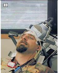
Repetitive Transcranial Magnetic Stimulation (rTMS) is a treatment for depression that was approved by the FDA in October of 2008 (1). Repetitive TMS involves a device (pictured right), which is noninvasive, that excites the neurons in the brain. When this done over the left dorsolateral prefrontal cortex (an area of the brain supposedly less active in depressed patients), brain activity increases. The major selling point is that it has very few side-effects compared to standard antidepressant treatment (most common effects are headache and tingling at the stimulation site).

The FDA approval of this device has been controversial (2). The initial study submitted to the FDA was rejected. The folks at Neurostar (the manufacturers of the device) did a post-hoc analysis of that data. They discovered that patients, who failed to respond to only 1 antidepressant, subsequently responded to rTMS greater than sham (27.3% versus 10.5%). Based on this analysis, the FDA approved rTMS for the treatment of MDD in patients who have failed only 1 antidepressant trial.

In this month's Archives of General Psychiatry, is an article titled "Daily Left Prefrontal Transcranial Magnetic Stimulation Therapy for Major Depressive Disorder" (3). This study was funded by the NIMH and is the first nonindustry funded multisite study of rTMS (though some of the researchers are paid consultants of the TMS manufacturer). It involved 190 people.

What supposedly separates this study from all others, is the sham treatment. One major criticism of the previous TMS research is that the sham treatment was not convincing enough to prevent unblinding (for example, sham did not cause scalp irritation or facial twitching). The researchers went to great lengths to develop a sham treatment which produced the similar physical sensations of rTMS to prevent unblinding.
(Click to Enlarge)


Unfortunately, approximately 50% of the active treatment group correctly guessed which treatment condition they were in. A full 66% of placebo participants correctly guessed their condition. In truth, the level of unblinding is not a whole lot different from standard antidepressant drug trials (since placebos are inactive). The patients, on average, were similar to the patients in the Neurostar post-hoc analysis. The participants failed 1.51 antidepressant trials. 
 
The primary outcome was remission, defined as a score of 3 or less on the HAM-D or 2 consecutive HAM-D scores less than 10 during phase 1 of the study. Phase 1 was three weeks in duration. Patients received rTMS once a day for 50 minutes (5 days a week).
(Click to Enlarge)
Unfortunately, the results were negative. During the three week period (the right side of the chart) only 6 patients (11%) met criteria for remission. The average drop in HAM-D score for active treatment was only 5 points (26 to 21). The researchers then extended the the length of phase 1 by two weeks. The number of patients who went into remission during this extension phase was 13 (14%). By increasing the length of the phase 1, they obtained statical significance. Sounds fishy to me, but at least they provide all the data.
 
Similar to the Neurostar analysis, those who did remit were less treatment resistance (i.e., failed only 1 antidepressant trial). The number needed to treat (NNT) was 12. That means, 12 people will need to be treated with rTMS before another person, who otherwise would have not remitted without intervention, finally does remit. That's not very good. However, that number is not far off from standard antidepressant drug trials.
 
Does rTMS have any practical value as a future treatment for depression? Based on these results, one will need to attend a 50 minutes session everyday (excluding weekends) for 3-5 weeks to see some sort of result. That is in stark contrast to attending psychotherapy 1-2 times a week or visiting a psychiatrist once every 4-6 weeks. As Daniel Carlat points out in his monthly report (1), each treatment session would cost approximately $400. Insurance companies do not currently cover this treatment (and probably never will). Moreover, the group of patients who did remit (i.e., those who failed only 1 antidepressant trial) is not very marketable. Odds are they will try a second antidepressant instead. According to the Star-D results, the odds of improvement are 30% on a second antidepressant compared to 14% of rTMS. Presently, rTMS just does not make economic sense.

ResearchBlogging.org

George MS, Lisanby SH, Avery D, McDonald WM, Durkalski V, Pavlicova M, Anderson B, Nahas Z, Bulow P, Zarkowski P, Holtzheimer PE 3rd, Schwartz T, & Sackeim HA (2010). Daily left prefrontal transcranial magnetic stimulation therapy for major depressive disorder: a sham-controlled randomized trial. Archives of general psychiatry, 67 (5), 507-16 PMID: 20439832

Tuesday, May 4, 2010

Psychosis Among Substance Users

Psychosis among drug users is quite common. Often, it is difficult to determine which came first, substance use or psychosis. Frequently, they co-occur. In cases where drugs are causally related (i.e., substance-induced psychosis), the condition is typically transient with a duration of 1 month or less. In rare cases, the length of psychosis can last longer. And in even rarer cases, symptoms such as hallucinations can be permanent.

Psychosis can be associated with the use many legal (e.g., alcohol) or illicit substance such as stimulants (amphetamines and cocaine), cannabis, and hallucinogens. An article by Thirthalli and Benegal (1) reviews the evidence that these drugs can cause psychosis in nonpsychotic persons. 

Alcohol
The neurochemical effects of alcohol are complex. "Common knowledge" states that alcohol acts in a similar fashion to other sedatives (e.g., diazepam). In other words, it is an agonist of GABA receptors. In reality, the alcohol molecule is very simple. It has the ability to cross cell membranes (e.g., blood-brain barrier) easily and can exert its effects on the brain within minutes. Alcohol also influences the phospholipid bilayer that make up cell membranes. This ability has a widespread impact on normal cell functions and also enables alcohol to modify the action of many neurotransmitter systems, such glutamate, dopamine, and norepinephrine in addition to GABA.

Alcohol-induced psychosis can occur during different drug states such intoxication or withdrawal (e.g., delirium tremens, alcoholic hallucinosis). In general, the risk of psychosis is two-fold greater than in the general adult population.

Stimulants (click to enlarge picture; 2)
Cocaine and Amphetamines are widely known to lead to psychosis. The psychosis produced by both cocaine and amphetamines is similar to schizophrenia. The risk of psychosis from amphetamine use is quite high; greater than 70% in chronic users. Users who develop first episode psychosis use an average of 20 times years. Psychosis typically lasts for the duration the drug is in the system. However, it can last more than a month in more severe cases.

For cocaine, typically 50% of chronic users experience paranoia and hallucinations. Cocaine-induced psychosis has a stereotypical form; Users believe that their drug use is being watched and that they are being followed. This paranoia is typically accompanied by hallucinations. Cocaine-induced psychosis also shows sensitization; that is, psychosis becomes more severe and occurs more rapidly with continued use. Unlike amphetamine-induced psychosis, cocaine induced-persistent psychosis is very rare.

Cocaine has the highest affinity and binds most strongly to the serotonin (5HT)reuptake pump, followed by the dopamine (DA) reuptake pump, then the norepinephrine (NE) reuptake pump (FYI-Effexor is cocaine with a PG-rating; effexor's affinities are for serotonin, then NE, and then DA). Contrary to popular belief, amphetamines do not act by blocking the dopamine reuptake pump. Amphetamines are indirect agnonist of the catecholaminergic systems (i.e., dopamine and norepinephrine). First, amphetamines go inside the neuron and release both DA and NE from their vesicles into the cell cytoplasm (i.e., catecholamins are released inside the neurone). Second, The catecholamines are subsequently transported outside of the neuron by a reversal of the reuptake pumps. This results in a MASSIVE increase in synaptic DA and NE. Lastly, at higher doses, amphetamines inhibit catecholamine metabolism, leading to even higher concentrations in the synapse. 

Cannabis
There has been a boom in the current research of cannabis and psychosis. There appears to be a temporal correlation between early cannabis use and onset of schizophrenia. This association is stronger than for any other substance. Why the association exists is unclear. In general, cannabis has the same risk of inducing psychosis as alcohol (i.e., two-fold).

There are two cannabinoid receptors in the human body: CB1 and CB2. The CB2 receptor is not expressed in the brain, and is primarily found in the immune system. The CB1 receptor is typically found in the basal ganglia, cerebellum, hippocampus, and the cortex. CB1 receptors exist on the axon terminal instead of the post-synaptic cell. In others words, CB1 receptors are autoreceptors that can inhibit the release of many different neurotransmitters.

Hallucinogens
Many different drugs fall under this category: mushrooms, peyote buttons, and LSD, for example. Many hallucinogenic drugs are either synthesized by plants or are based on plant-derived compounds. The main active compound in peyote is mescaline, while psilocin in found mushrooms. LSD is actually a synthetic compound, but is based on a fungal alkaloid taken from ergot.

Hallucinogenic compounds have a catecholamine-like structure (most are similar in shape to serotonin). Hallucinogens are primarily 5-HT2a receptor agonists. While these drugs do not lead to dependence, withdrawal, or cravings, they still can lead to serious problems for some users. Some people experience acute anxiety or panic attacks in responses to the drugs' hallucinogenic effects. There is a disorder known as hallucinogen persisting perception disorder (HPPD), which is the fancy name for "flashbacks." The most severe reactions, of course, are psychotic breakdowns. However, similar to the above mentioned drugs, psychosis is typically transient. Most prolonged episodes of hallucinogen-induced psychosis involve individuals who have already been diagnosed with a psychotic disorder or who have manifested prepsychotic (e.g., prodromal) symptoms before taking these drugs.

Treatment
In most cases, substance induced psychosis does not need medical treatment per se. It usually disappears when the drug's affects are gone. However, there are cases when treatment is necessary. Alcohol dependent individuals in withdrawal do need medical treatment because alcohol withdrawal can be lethal. Typically, sedatives (benzodiazepines) are the drugs of choice. For either cocaine or amphetamine induced psychosis, first or second generation antipsychotics are the drugs of choice because of their potent D2 receptor antagonism. In general, it is common for anyone presenting with psychotic symptoms to be prescribed antipsychotics. Occasionally, patients who are addicted to amphetamines will be prescribed antipsychotics in hopes that they will reduce the risk of subsequent psychosis or reduce euphoria, making the drug less reinforcing. Drugs that antagonize the 5HT2a receptor such as risperidone (or any second generation antipsychotic), ketanserin, and ritanserin have been shown to reverse hallucinogenic-induced psychosis.

In rare cases when psychosis persists, there are a few things to consider, such as, is their another cause for the psychosis? For example, does the person have schizophrenia or is there a physical cause for the psychosis (e.g., tumor, metabolic, etc)? Often times, doctors not knowing what to do, will double down on the antipsychotics. This is unlikely to work because the mechanism of action (D2 or 5HT2a blockade) does not necessarily increase with the addition of a second drug. Simply upping the does of the current drug should suffice. Side effects, however, are always additive. Antipsychotics with higher affinities for both D2 and 5HT2a receptors are preferable (e.g., risperidone).

Risk Factors
The risk factors for substance-induced psychosis are similar across all substances. Pre-morbid psychiatric history or a family history of schizophrenia put an individual at risk. Also, the longer a substance is consumed and the larger the quantities consumed are also risk factors. Polysubstance use or consuming drugs that contain other compounds increases the risk of psychosis too. Unfortunately, the research into the neurobiological and genetic underpinnings is substance-related psychosis are quite poor. A useful theory for substance-related psychosis which could lead to better acute treatment is lacking.

ResearchBlogging.org

Thirthalli, J & Benegal, V. (2006). Psychosis Among Substance Users Current Opinion in Psychiatry

Monday, May 3, 2010

"Anatomy of an Epidemic" or The Same Story Told Over and Over Again

On Salon.com, there is an interview with Robert Whitaker, the author of Mad in America, about his new book titled "Anatomy of an Epidemic: Magic Bullets, Psychiatric Drugs, and the Astonishing Rise of Mental Illness in America."

The article/interview at Salon is devoid of any context:
"In the past few months, the perennial controversy over psychiatric drug use has been growing considerably more heated. A January study showed a negligible difference between antidepressants and placebos in treating all but the severest cases of depression. The study became the subject of a Newsweek cover story, and the value of psychiatric drugs has recently been debated in the pages of the New Yorker, the New York Times and Salon...The timing of Robert Whitaker’s "Anatomy of an Epidemic," a comprehensive and highly readable history of psychiatry in the United States, couldn’t be better."
That study in JAMA is hardly a first (1, 2, 3), nor is Mr. Whitaker's book a first (4, 5, 6, and anything written by Peter Breggin). Timing couldn't be better? Sure, if by timing you mean, when can one cash in on anti-psychiatry sentiment.

I have not read this book. I did read his previous book, Mad in America, which surveyed the history of the treatment of mental illness in American and the rise of psychiatric drugs. Based on that book, which was sensationalistic and misrepresented research, I think I know what to expect in his new book. My focus, instead, will be on his interview. Comments in red.
"Psychiatric drug use is a notoriously tough subject for writers, because of all the contradictory research. Why wade into it? In 1998, I was writing a series for the Boston Globe on abuse of psychiatric patients in research settings. I came across the World Health Organization’s outcomes study for schizophrenia patients, and found that outcomes were better for poor countries of the world -- like India, Colombia, Nigeria -- than for the rich countries. And I was startled to find that only a small percentage of patients in those countries were medicated. I also discovered that the number of people on disability for mental illness in this country has tripled over the last 20 years..."
 He doesn't provide an actual number for "only a small percentage of patients." However, according to the Worldwide-Schizophrenia Outpatient Health Outcomes (W-SOHO;7), which surveyed Europe, Asia, Africa, and Latin America, it revealed that a not so small 40% of patients where on antipsychotic medications. The real differences between rich and poor counties is that patients in developing countries were primarily on first generation antipsychotics (60%) and on monotherapy (75%). The reasons for outcome differences are many.
"...If our psychiatric drugs are effective at preventing mental illness, I thought, why are we getting so many people unable to work?"
I don't think anyone of real importance claimed that psychiatric drugs were aimed at "preventing mental illness." Again, there is no context. For example, one explanation for the increase of those on disability is that entitlement programs have increased their coverage over the years to include those with mental illnesses.
"What's so risky about Ritalin? For one, a significant percentage -- between 10 and 25 percent -- of kids prescribed medication for ADHD will have a manic episode or psychotic episode and deteriorate in such a way that they’re diagnosed with bipolar disorder..."
He is misrepresenting the data. That study (8), examined the comorbidity of ADHD and mania, not the association of stimulant medication use and risk of mania. It's quite possible that those who eventually develop bipolar disorder have an ADHD appearance during childhood.
"But if these studies are so groundbreaking, why have they gone unreported in the media? Because the NIMH didn’t announce it. Just as they didn’t announce the 2007 outcome study for schizophrenia patients. In that study, the recovery rate was 40 percent for those off meds, but only 5 percent for those on meds. I checked all the NIMH press releases for 2007, and found no release on this study. I found no announcement of it in any American Psychiatric Association publication or textbook. Not a single newspaper published an account of the study. And that’s because the psychiatric establishment -- the NIMH, the APA, even the National Alliance on Mental Illness, an advocacy organization -- did not put out any press release about it or try to alert the media in any way."
Either he's lying or had a lobotomy. I have reviewed that study as well as it's follow-up (9). Here is what the researchers actually discovered :
"Looking at it from a different viewpoint, the data suggests that schizophrenia patients with good prognostic features, with better premorbid developmental achievements and with more favorable personality characteristics are the subgroup more likely to stay off antipsychotics for a prolonged period"
That's why the NIMH, APA, and NAMI didn't sound the alarm about that supposed discovery. Here's another way to highlight the absurdity of his conclusion: Two women have been diagnosed with breast cancer. One has a strong family history of breast cancer and tested positive for the BRCA gene (i.e., she has a poor prognosis). The other woman has no family history nor the BRCA gene. Both were treated with chemo. One recovered quickly stopped treatment. The other woman, while still being treated, died. The chemotherapy killed her, right?

 I could continue, but what's the point. His book has one intended audience: People who already hate psychiatry. If one cares to fully fact check the entire interview, you'll find one misleading statement after another. Whitaker is biased and has no scruples about misrepresenting facts and data to suit his narrative.

He is no different than the very industry that he is attacking.  

Tuesday, April 27, 2010

R-Rated Movies Aren't Bad for Your Kids...R They?

In the May issue of the Journal of Studies on Alcohol and Drugs is an article titled "Parental R-Rated Movie Restriction and Early-Onset Alcohol Use." (1) Without reading the study, one can guess the reported result : the more R-rated movies that youths watch, the higher the frequency of early-onset alcohol use.

This study was funded by the National Cancer Institute and National Institute on Alcohol Abuse and Alcoholism. Remember what the Last Psychiatrist says: What do researchers want to be true?

First comes the filter that will aid our interpretation of the results:
"TODAY’S YOUTH HAVE UNPRECEDENTED ACCESS to entertainment media (Roberts et al., 2005), and longitudinal research has linked various forms of entertainment media with aggressive behavior (Bushman and Anderson, 2001), sexual behavior (Collins et al., 2004), and tobacco use (Sargent, 2005). Indeed, the available research evidence has led the National Cancer Institute (2008) to declare a causal relation between exposure to movie smoking and youth smoking initiation."
In the words of Mr. Mackey, "Mm..R-rated movies are bad...mmkay."

The study was done by survey. 3,577 youths where chosen out of 4,655 (grades 5-8) because they reported to never have drunken alcohol without the knowledge of their parents. Also at baseline assessment the youths rated how restrictive their parents were in allowing them to view R-rate movies.

Statistics 101: Very boring, but very important to interpret these results.

This study is correlational. This results are known as the standardized coefficient. This can vary from +1 (indicating a perfect positive relationship), to zero (indicating no relationship), all the way down to -1 (indicating a perfect negative relationship, that is, when the frequency of a factor goes up, the other factor goes down). As a rule of thumb, standardized coefficients between .00 and .30 are considered weak, between .30 and .70 are moderate, and between .70 and 1.00 are considered strong.

The primary meaning of standardized coefficients lies in the amount of variation in one variable that is accounted for by the variable with which it is correlated. To obtain this information, you square the standardized coefficient (e.g., .30 x.30). This number is called the coefficient of determination. Then you multiply the coefficient by determination 100. This proportion of variance indicates the percentage of variance. The coefficient of determination is the primary information measure. Correlation coefficients of .30 account for about 10 percent of the variance (.30x.30 =.90x100= 9%).

Here are the results as reported in the abstract, that is, the information they want you to know:
"The sample included 2,406 baseline never-drinkers who were surveyed at follow-up, of whom 14.8% had initiated alcohol use. At baseline, 20% reported never being allowed to watch R movies, and 21% reported being allowed all the time. Adolescents allowed to watch R-rated movies had higher rates of alcohol initiation (2.9% initiation among never allowed, 12.5% once in a while, 18.8% sometimes, and 24.4% all the time). Controlling for sociodemographics, personality characteristics, and authoritative parenting style, the adjusted odds ratios for initiating alcohol use were 3.0 (95% CI [1.7-5.1]) for those once in a while allowed, 3.3 [1.9, 5.6] for those sometimes allowed, and 3.5 [2.0, 6.0] for those always allowed to watch R-rated movies. Alcohol initiation was more likely if R-rated movie restriction relaxed over time; tightening of restriction had a protective effect (p < .001)."
With odds-ratios, 1.0 represent the prevalence of something (e.g., drinking behavior) that already exists in the population. Anything above that represents an increase in prevalence. For example, in the study, it's reported that youths who have peers who drink have an OR of 5.7, meaning that there is over a 400% increase in the incidence of early on-set drinking when one's peers start drinking early (peer pressure). Kids allowed to view R-rated content once in a while showed a 200% increase, those allowed sometimes saw a 230% increase, and those allowed all the time saw a 250% increase.

While these numbers are quite high, this tells us nothing about the relationship strength. Remember multiple variables can lead to this result. Here is a graph of the correlation coefficients:
Exposure to R-rate movies has a standardized coefficient of 0.10. This relationship is weak. Using the formula above, the amount of variance in behavior attributed to viewing R-rated content is only 1%. 

So why are youths, who are exposed to R-rated content, 2 times more likely to try alcohol at an earlier age?

Here is how the authors explain it:
"Youth who say that their parents allow them to watch R movies see more R movies and, therefore, more depictions of alcohol use...thus, the mechanism could be social influence via modeling of positive depictions of alcohol use."
Wrong!

Exposure to R-rated movies various with onset of early alcohol use by 1%. That means 99% of the variance is explained by other reasons (e.g., peer pressure!) While it is true that this tiny relationship is statistically significant, it has no practical significance what so ever.

But who cares about the actual science, politicians and other nanny state advocates have their sound bite,
"The research to date suggests that keeping kids from R-rated movies can help keep them from drinking, smoking and doing a lot of other things that parents don't want them to do, Sargent said." (2)
If only it were that simple.

This post was chosen as an Editor's Selection for ResearchBlogging.org

Tanski SE, Cin SD, Stoolmiller M, & Sargent JD (2010). Parental R-rated movie restriction and early-onset alcohol use. Journal of studies on alcohol and drugs, 71 (3), 452-9 PMID: 20409440

Thursday, April 22, 2010

Do Antidepressants Enhance Stroke Recovery?

A few months ago, in the February issue of Archives of General Psychiatry appeared this study (1) title, "Escitalopram and Enhancement of Cognitive Recovery Following Stroke."

The design was simple: a placebo group, an escitalopram group, and a problem solving therapy group (a manual based therapy to treat depression in the medically ill). No patients in this group met diagnostic criteria for Major Depression (so why the PST group?) according to the Hamilton Scale for Depression (HAM-D). Patients were administered the following neuropsychological tests at baseline and post-treatment:

"-The Repeatable Battery for the Assessment of Neuropsychological Status (RBANS). This 25- to 30-minute battery assesses functioning in 5 domains (immediate memory, visuospatial/constructional, language, attention, and delayed memory). These domains are evaluated by 12 individual subtests, including List Learning, List Recall, List Recognition, Story Memory, Story Recall, Figure Copy, Figure Recall, Line Orientation, Picture Naming, Semantic Fluency, Digit Span, and Coding. This battery provides age-corrected norms for overall performance (total scale score) and scores in each domain."

(Number of Subjects in Each Treatment Group)

Here is what the researchers discovered: "We found a difference among the 3 treatment groups in change in RBANS total score (P<.01) and RBANS delayed memory score (P<.01)." They also found a significant result for immediate memory too, but for reasons beyond my knowledge (crack-cocaine?) they neglected to report this result. Test scores are below.

(Click to Enlarge)

The authors then engaged in a trite discussion about how escitalopram led to an improvement in memory and how serotonin theoretically affects memory, and blah, blah, blah.

On the surface, it does appear that escitalopram led to better outcomes: For the Total Score (a global measure of cognition) the escitalopram improved by 9.1 points compared to 5.7 for placebo; for delayed memory the escitalopram group improved by 12.4 points versus 5.9 for placebo, and for immediate memory (i.e., learning) the escitalopram group improved by 11.7 points versus 7.4 versus placebo.

However, look at the post-treatment final scores for both groups: total score: 89.8 (escitalopram) versus 91 (placebo); delayed memory 96.6 (escitalopram) versus 94.2 (placebo); and immediate memory 95.1 (escitalopram) versus 98.5 (placebo). Essentially, both groups post-treatment performance was nearly identical. The reason why the escitalopram group showed a larger magnitude in change was because that group had lower baseline scores.

The authors did point out that there was not statistical different between any of the baseline scores; however, that's irrelevant. Neuropsychological test performance scores are classified as either average, low average, mild deficit, moderate deficit, or severe deficit. Many of the escitalopram group baseline scores where in the mild-deficit range while the placebo group scores were in the low-average range. Functionally, that kind of difference is significant*.

Another factor not discussed is the rate of recovery in stroke patients. There is a pretty well delineated recovery curve for patients with mild-to-moderate strokes. Most recovery occurs in the first 6 months, followed by a slower recovery over the next 1-2 years. Those with more severe deficits will show greater initial recovery and then plateau at a level similar to the less sever group. This is why the groups matched on post-treatment assessment scores.

*It's actually more complicated than that. Scores actually fall within a certain range. So a person could perform in the mild-deficit range at point then in the low-average range at another point. So it's quite possible that these results reflect the error in the psychological instrument rather than a benefit from escitalopram.

Either way, I'm skeptical that escitalopram actually helped these people.

ResearchBlogging.org

Jorge RE, Acion L, Moser D, Adams HP Jr, & Robinson RG (2010). Escitalopram and enhancement of cognitive recovery following stroke. Archives of general psychiatry, 67 (2), 187-96 PMID: 20124118

Monday, March 15, 2010

WARNING: Chantix Causes Suicide, Herpes, and Nocturnal Vaginal Itch Syndrome

Here is a brief tale of an American dream deferred...
"Tina Hurst is a suburban mother and a manager at a Fortune 500 company. Her life was great, but she had one dark secret...'I was a closet smoker,' Hurst said."  
Here is where the plot thickens...
"Her doctor prescribed a drug called Chantix to help her quit. Seven million Americans have taken it. Hurst says she took Chantix for two months and it worked great."
Then, tragedy struck...
"But when she stopped, 'I totally went off the deep end,' she said. Hurst says she started walking around the house in circles, crying and hallucinating. 'I thought somebody died. And I was freaked out,' Hurst said." 
But, it wasn't over just yet...
"An ambulance rushed Hurst to a hospital where records indicate she was very agitated and had to be restrained. She spent four days in a psych unit. Prior to this, Hurst said she never had a history of anxiety, depression or any mental problems. She thinks it was caused by Chantix. Her doctor's diagnosis? "Substance-induced psychotic disorder." (1
No, this isn't the plot of Gothika (2), this is an account of a woman who claims that she was adversely affected by Pfizer's anti-smoking drug Chantix (varenicline). Stories like this are common place now. Since the drug has come to market, it has racked up some 10,102 reports in the FDA's adverse events database (3).

Normally, I'm all for psychoactive drugs causing harm to people. However, I think a skeptical approach should be taken when it comes to this drug.

The most widely heard side-effect of Chantix is that it causes suicidality. For the sake of argument, let's forget that suicidality is a very complex and multifaceted human condition; we're going to pretend that this little pill can lead an otherwise normal and healthy person to want to kill himself (4).

First, let's examine the neurobiological reasons why this might happen...oh, wait a minute, there are no widely accepted (or proposed?) mechanisms of how this happens, just like there is no proposed mechanism for the supposed anti-depressant induced mania phenomenon (5) or the anti-depressant induced suicidality phenomenon.

What we do have is a temporal association (6). That is, event B happens around, near by, within some sort of proximity of event A. Once that happens, our minds automatically link events A and B together, and we have causation! No need for scientific investigation. Just turn on your computer, sign onto word press and start blogging about how "Chantix Causes Traffic Accident, Passengers Almost Die."

Here's why I am skeptical of the Chantix-suicidality link: smoking is associated with psychiatric illness (8, 9, 10). Specifically, individuals who smoke and have a comorbid psychiatric (i.e., Axis I or II) disorder make up 7% of the population, yet they consume 34% of all cigarettes smoked in the United States. Moreover, cigarette use is associated with most major psychiatric disorders. This means that 7% of 45 million smokers are mentally ill. That's a lot of people who are already at risk for suicide, regardless of whether they use Chantix or not.

Here is another interesting piece of research:
"This study examined the relation between smoking and suicide, controlling for various confounders...We found a positive, dose-related association between smoking and suicide among White men. Although inference about causality is not justified, our findings indicate that the smoking-suicide connection is not entirely due to the greater tendency among smokers to be unmarried, to be sedentary, to drink heavily, or to develop cancers." (11
I almost forgot about this study:
RESULTS: Current daily smoking, but not past smoking, predicted the subsequent occurrence of suicidal thoughts or attempt, independent of prior depression and substance use disorders (adjusted odds ratio, 1.82; 95% confidence interval, 1.22-2.69). Additionally, current daily smoking, but not past smoking, predicted the subsequent occurrence of suicidal thoughts or attempt, adjusting for suicidal predisposition, indicated by prior suicidality, and controlling for prior psychiatric disorders (adjusted odds ratio, 1.74; 95% confidence interval, 1.17-2.54).  (12)
Anybody ever abruptly stop their anti-depressant medication? Bad stuff happens, right? Here is a pretty picture from a study in the September 2003 issues of PENIS er, PNAS (13).


Here is a comparison of a smoker and nonsmoker and the amount of monoamine oxidase throughout their bodies. Smoking lowers the amount of this enzyme, which breaks down the catecholamines (5HT, DA, NE). Without this enzyme, the levels of these neurotransmitters rises. This is how the class of anti-depressants known as monoamine oxidase inhibitors are supposed to work.

Here are the instructions for how a patient is supposed to use Chantix:

 That is, they quit smoking one week after starting Chantix. Since Chantix is a partial-nicotinic agonist and not an MAOI, this could have the same effect as abruptly stopping your anti-depressant medication.

What if the instructions read like this instead. "Patients should be instructed to set a date to quit smoking and to stop their antidepressant medication and to initiate CHANTIX treatment one week before the quit date." Would you still take it?

Earlier, I mentioned the FDA's adverse events database. This is a system, where if an adverse reaction to a drug is suspected, it can be reported.

Here is a link (14) to the FDA's adverse events database on those who quit smoking cold turkey.

You'll notice that there is no real link because there is no actual database for this. The data that is collected by the FDA's actual database is biased, there are too many confounds that are not accounted for.

What about actual research? If you search PubMed for the terms "varenicline" and "suicide" you get 19 hits. Only three are actual prospective studies that looked at the matter (15, 16). The first study only had 1 suicide, but the patent's psychiatric background is unknown since the study is not complete. In the second study,
"There was no evidence that varenicline was associated with an increased risk of depression or suicidal thoughts."
The third study uses an adverse events reporting paradigm (17):
"All patients with suicidal events either had a past medical history of psychiatric illness prior to starting varenicline and/or a precipitating factor for the event. Clinicians should closely monitor patients with pre-existing psychiatric illness who are taking varenicline."
I'm not defending Chantix or Pfizer; over 10,000 reported adverse events for a drug that has been on the market for less that 4 years is a lot. Also, I have no explanation for how seemingly normal people such as Ms. Hurst have such horrible reactions. But, let's not always play the "drug companies are evil" card either. Chantix is the most effective smoking cessation drug on the market. It actually has helped 2 or 3 people.

Just remember, things are not always as they seem; do your own research; and don't trust the shit you read on blogs!

Thursday, February 25, 2010

Is the Clinical Significance Criterion Significant?

The draft version of DSM-V: Revenge of the Fallen has been online for a few weeks (1) and much has already been written about it (1, 2, 3, 4). Much focus has been on what is "new" and what is "gone." One feature that is shared by the majority of DSM diagnoses, the "clinical significance" criterion, might be on its way out. Typically this criterion reads "The symptoms cause clinically significant distress or impairment in social, occupational, or other important areas of functioning." The general rule being, if the person does not satisfy this criterion, a diagnosis probably should not be made.

This criterion is unique to the DSM-IV and is not found in the earlier versions of the text. The stated reason for adding it to the DSM was to
"establish the threshold for the diagnosis of a disorder in those situations in which the symptomatic presentation by itself (particularly in its milder forms) is not inherently pathological and may be encountered in individuals for whom a diagnosis of mental disorder would be inappropriate."  
Since mental health disorders are made by subjective analysis (often referred to as clinical judgment), does the addition of this criterion aid in the diagnostic process?

Not according to Wakefield et. al who published an article in the January 2010 issue of the American Journal of Psychiatry (5). In the article, titled, "Does the DSM-IV Clinical Significance Criterion for Major Depression Reduce False Positives? Evidence From the National Comorbidity Survey Replication," the authors' reason why the criterion is ineffective, is because it is redundant.

Wakefield argues that "distress is common to both normal reactions (e.g., acute grief) and disordered conditions, 'since most of these symptoms are either intrinsically distressing or are almost invariably accompanied by distress about having the symptom."

In other words, it's highly unlikely that an individual will satisfy full diagnostic criteria for a disorder and not be distressed or impaired.

(Click to Enlarge)

In the results reproduced above, out 2,071 respondents who reported episodes of sadness, 1,254 (60.5%) met diagnostic criteria for major depressive disorder (MDD). Of those who did not meet full criteria for MDD (n=817 or 39.5%), 93.5% did satisfy the "clinically significant distress or impairment" criterion. This suggests that the criterion is a poor indicator of diagnostic status. This result agrees with other research (6). 

It's unlikely that any modification of this criterion, other than its deletion, will resolve this issue of redundancy. If the definition is narrowed, there will be more false negatives; if the definition is broadened, there will be more false positives. Actually, the whole idea of false negatives/positives for already highly arbitrary (i.e. not valid) diagnoses is quite humorous, but I digress...

ResearchBlogging.org

Wakefield, J., Schmitz, M., & Baer, J. (2010). Does the DSM-IV Clinical Significance Criterion for Major Depression Reduce False Positives? Evidence From the National Comorbidity Survey Replication American Journal of Psychiatry DOI: 10.1176/appi.ajp.2009.09040553

Wednesday, February 24, 2010

A Tale of Two Studies: Voxel-Based Lesion-Symptom Mapping

Brain imaging has contributed greatly to our understanding of the functional neuroataomy of the human brain. A lot these contributions have been blogged about by my bestest buddy Neuroskeptic (why don't you return my phone calls anymore!?). One of the more popular methods used to capture brain function is the functional magnetic resonance (fMRI). However, the results of fMRI studies are correlational and do not represent causation. There is another method, however, that "can identify regions, including white matter tracts, playing a causal role in a particular cognitive domain." This method is known as voxel-based lesion-symptom mapping (VLSM). A voxel is the three-dimensional analog of a pixel, and represents a volume of about 1 cubic millimeter. This method produces pretty images such as this one below.
A team of researchers from various important sounding universities published a study in this month's Proceedings of the National Academy of Sciences (PNAS; 1). In this issue of PNAS (pronounced penis), is an article titled "Distributed Neural System for General Intelligence Revealed by Lesion Mapping." The researchers created 3-D representations of the lesions of 241 subjects who had "single, focal, stable, chronic lesions of the brain." The subjects also had undergone neuropsychological testing, which included either the WAIS-R/WAIS-III.

The researchers were trying to discover where in the brain is general intelligence (often designated as "g"). Specifically,
"we address the question of whether g draws upon specific brain regions, as opposed to being correlated with global brain properties (such as total brain volume). Identifying such brain regions would help shed light on how g contributes to information processing and open the door to further exploration of its biological underpinnings, such as its emergence through evolution and development, and its alteration through psychiatric or neurological disease."
If "g" sounds like a highly abstract to concept to you, that's because it is. It's actually a really controversial concept within the field (2, 3). Below are the "g" loadings from this study.
The closer the color is to red, the closer that particular subtest loaded onto one of three g-related functions (i.e., verbal, spatial, working memory). The statistics of this study are admittedly over my head, since calculating g loadings require factor analysis. Since "g" is an abstraction, no actual number is presented for "g." Only how well a specific test loads onto "g" is provided.

What the researchers discovered should not be surprising to any biped mammal with working frontal lobes,

"One of the main findings that really struck us was that there was a distributed system here. Several brain regions, and the connections between them, were what was most important to general intelligence." (4)
More specifically,
"Statistically significant associations were found between g and damage to a remarkably circumscribed albeit distributed network in frontal and parietal cortex, critically including white matter association tracts and frontopolar cortex. We suggest that general intelligence draws on connections between regions that integrate verbal, visuospatial, working memory, and executive processes." (1)
"Statistically significant associations" is not same as "causal role." It's correlational. Still, nice sleight of hand.

What this group of geniuses is saying is that different brain functions are located in different parts of the brain, and when everything works in harmony, you have general intelligence.
"The researchers say the findings will open the door to further investigations about how the brain, intelligence, and environment all interact."
Open doors? That would mean that this research is original and ground breaking. It's not. In fact, in the March 2009 issue of Neuron (5) appeared this study, "Lesion Mapping of Cognitive Abilities Linked to Intelligence." Here is the press release (6). In this study, there were 241 patients with "single, focal, stable, chronic lesions of the brain," who had their lesions mapped and were also administered either the WAIS-R/WAIS-III. Also, the researchers are the same in both studies.

This study also found that performance on these (same) tests mapped primarily onto the frontal and parietal lobes.

The main difference is that the former study examined the anatomical location of intelligence in general, while the latter examine the anatomical location of general intelligence.

"So, what's the difference smart ass!?"

It depends on who you ask. Some say there is no difference, while others say there is a difference. At this point in the debate, however, we're engaging in mental masturbation (which is equally satisfying, plus people don't stare when you do it on the bus).

What I've been trying to figure out is if this counts as a duplicate publication? Sure, this doesn't have the far reaching consequences of these douche baggers (7, 8). There is a slight theoretical difference, the results are essentially identical. Curiously, in the most recent study, there is no citation to the other study. You'd think that the researchers want other people to read both of their studies.

Perhaps I'm looking too much into this. Or, perhaps I just enjoy mental masturbation...

ResearchBlogging.org

Gläscher J, Rudrauf D, Colom R, Paul LK, Tranel D, Damasio H, & Adolphs R (2010). Distributed neural system for general intelligence revealed by lesion mapping. Proceedings of the National Academy of Sciences of the United States of America PMID: 20176936

Gläscher J, Tranel D, Paul LK, Rudrauf D, Rorden C, Hornaday A, Grabowski T, Damasio H, & Adolphs R (2009). Lesion mapping of cognitive abilities linked to intelligence. Neuron, 61 (5), 681-91 PMID: 19285465

Wednesday, February 3, 2010

Brodmann's Map 100 Years Later

Brodmann's map. Anyone who has taken a course in basic neuroanatomy has been exposed to his roadmap of the cerebral cortex.

In this month's Nature Reviews Neuroscience, Zilles and Amunts (1) dedicated an article to Korbinian Brodmann and his map, celebrating its 100th anniversary (Brodmann's original work was published in 1909).

First, a little background. Brodmann's original map contains 52 areas; however, areas 12-16 and 48-51 are only found in nonhuman primate brains, so only 43 areas are actually labeled. How Brodmann constructed his "map" is quite complicated. He made numerous razor thin, horizontal slices of human brains. He then stained the cell bodies within those slices and attributed a number to an area if it was cytoarchitectonically distinct from its neighboring areas of the cortex.

Many others followed Brodmann's work with maps of their own. According to the article,
"During the next three decades, Otfried Foerster, Alfred Walter Campbell, Grafton Elliott Smith, Constantin Freiherr von Economo and Georg N. Koskinas argued for localizable anatomical and functional correlation and the segregation of cortical entities"
Many of those names may be new to you, which highlight how influential Brodmann's work has been. The reason there are many different "maps" is because brain mapping is not an exact science. Trying to differentiate the cortex based on brain architecture can produce profoundly different results, depending on the staining technique that is used and on the researcher's subjectivity.
"The Vogts used myelin-stained histological sections to study brain architecture (that is, myeloarchitecture). Their myeloarchitectonic map has many more areas (a total of 200) than that of Brodmann, because the Vogts further subdivided the Brodmann areas on the basis of the regionally more differentiated architecture of intracortical nerve fibres."
Below is a comparison of the various "maps" that have been produced since Brodmann's work in 1909.
(click to enlarge)
Differences between all these brain maps are apparent. However, there is also considerable overlap, suggesting that there is some degree of observer independence, reproducibility, and objectivity to the process.

A little historical note for anyone who was forced to memorize all those Brodmann areas, but was hampered by its apparent lack of logic (areas 1,2,3, start in the mid-lateral areas, while the remaining numbers are distributed in a quasi-random order). Each area number was assigned based on the order in which he prepared a slide, hence the apparent randomness of number assignment.

In his time, testing whether each "area" was correlated to a specific function was quite difficult. Over time, as other "maps" were published and his original became criticized for lack of objectivity, his map fell out of fashion. That is until the 1980's, when various brain imaging techniques were developed. Being able to image a live human during the performance of a specific task, it became possible to associate functional data with cytoarchitectual data. It was Brodmann's map that become apart of many of the first software and sterotaxic atlases for these machines.

Brodmann's work helped to revolutionize modern neuroscience. While many other maps have followed Brodmann's, and even though contemporary research has shown that "his map is incomplete or even wrong in some of the brain regions," many of the areas do correlate very well with various functional areas of the cortex, which is why his work still has relevance 100 years later.

This post was chosen as an Editor's Selection for ResearchBlogging.org

Zilles K, & Amunts K (2010). Centenary of Brodmann's map - conception and fate. Nature reviews. Neuroscience, 11 (2), 139-45 PMID: 20046193